CT
Scans in Art Work Appraisal
by Dr Marc Ghysels
October 25, 2005
| please click here for the French version |
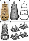
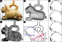
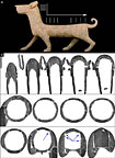
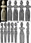
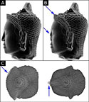
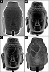
(click on small images below for large images with captions)
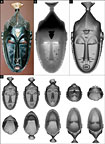
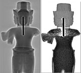
Since the late 1970s, CT scanners have probed patients’ bodies to help identify the causes of their illnesses. Just as the technique of computed tomography imaging revolutionized the practice of medical diagnosis in its time, its contemporary use in the art world could ultimately change the way some works are appraised. The quality and reliability of the images produced by a CT scanner – also called a computed axial tomography scanner or a CAT scan – literally “undress” the art work and reveal its internal structure.
The CT scanner, or “CT”, as it is more briefly known, provides a more accurate measurement of the density of the component parts of the object under examination, thereby dissociating parts that are usually merged on a conventional X-ray film. It therefore has its place among the various scientific disciplines used to clarify the history of art works: manufacturing techniques, initial functions, later uses, preservation, etc.
In conventional radiology, the X-ray beam projects onto the film the accumulated shadows of the component parts of the object it goes through. Low-density areas are completely masked by the shadow of denser parts. CT avoids this drawback by enabling each part to be viewed separately. The principle is to record a series of “slices” or sections of the object. The images are recorded in digital format and special image processing software is used to construct sections on any spatial plane desired. The sections can be combined to give a global view of the object and the transparency of the component parts can be modified at will. These operations reveal valuable information about the object’s background:
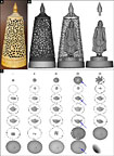
- manufacturing techniques,
showing for example, whether pottery was modeled from a clay coil, turned
on a wheel or stamped,
- natural damage, such as oxidation, erosion, or cracking,
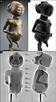
- repairs: impregnation, infiltration, gluing of chips or fragments, etc.,
- restoration: a clear and accurate reconstruction of the damaged parts,
- even the tricks used in assembling the piece.
The technique of CT scanning, when combined with a pertinent interpretation of the images obtained, is a powerful diagnostic tool which can provide proof of the inner state of an art work. Although it can help establish the history of the piece, when used in conjunction with other techniques of observation and analysis, it is not a dating test.
Like all sophisticated equipment, the CT has its limits. It was designed principally to examine the human body so the three-dimensional objects scanned must not exceed a diameter of 50 cm or the weight of a man. Although a medical CT can handle most materials, an industrial CT should be used for metal items.
CT scanning of antiquities is not recent: in 1979, the year when the Nobel Prize for Medicine was awarded to Allan M. Cormack and Sir Godfrey N. Hounsfield for inventing the CT scanner, Dr Derek Harwood-Nash published the first article on his use of a CT to study an Egyptian mummy. He was already fully aware of the advantages that this essentially non-destructive technique had to offer scientific fields such as Egyptology, Paleontology and Archeology.
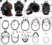
Since then, precisely because of its non-destructive nature, CT scanning has been widely used to examine objects from the past, as it leaves them intact for future generations. For example, a CT scan of an Egyptian sarcophagus can pinpoint the ceramic or stone scarab hidden in the mummy’s thorax and give a precise view of the underside. Any hieroglyphs engraved there can then be photographed, enabling an Egyptologist to decipher the history of the deceased without violating the sarcophagus.
Detailed reports of other applications in the cultural heritage field have been published in the press, in specialized magazines or on the Internet: CT scans have assisted in the study of stringed instruments (violins, cellos and guitars), archeological pottery vases, antique glasses, wooden and ivory netsuke, real and fake fossils, and dinosaur eggs. More recently, CT scanning has proved useful in making models of ancient musical instruments, identifying art works for insurance purposes, and making three-dimensional polymer reproductions of art works (stereolithography).
The value of CT for art works lies in the fact that each particle of material making up an object has a measurable density, which differs to some extent from the adjacent particles, because the composition of non-synthetic materials is seldom homogeneous. The density of these particles remains perfectly stable over time because it is mainly dependent on the concentration and average atomic weight of its atoms. When a physical or chemical change occurs in the object it will show up unfailingly as a change in the density of the material. For example, oxidation will cause a lowering in the density of the oxidized material in relation to the non-oxidized particles.
Since it is designed to measure the density of matter, the CT is particularly appropriate for detecting any change in the state of a material. The result of the examination is usually presented as an image in which the densest particles appear dark grey or black, while those of lesser density are of a lighter grey. Unfortunately, these images, displayed in a scale of greys, do not always speak for themselves! They must be interpreted, just as, in the medical field, a radiologist is needed to interpret a scan of the spinal column, for instance, to see whether or not the vertebrae are fractured. The quality of this interpretation obviously depends on several criteria such as the quality of the CT, its initial calibration, the adjustment of the parameters used for the examination, and the type of algorithm used to construct the images.
Apart from its non-destructive character, the CT has another substantial advantage over other scientific tests in that it examines the entire object rather than just a sample.
When used under optimal
conditions, its resolution power, that is the minimal diameter of a particle
of material for which the CT can measure a density value, is about 0.05
mm. This resolution power is therefore largely sufficient to detect, for
instance, a crack in a wooden, ivory, pottery or stone sculpture, even
if the crack is invisible to the naked eye and does not show up on a conventional
X-ray film.